Foot Muscles Mri. Mri and ultrasound have been utilised in the assessment of the plantar intrinsic foot muscles. Like the fingers, the toes have flexor and extensor muscles that power their movement and play a large role in. Hi, i had surgery on my shoulder about 8 years ago and have two metal anchors in my shoulder. Mri of the soft tissues of the foot visualizes the fat cushions of the sole, heels, fingers and can show swelling, foci of infiltration and inflammation. However, to establish a relationship between intrinsic muscle weakness and foot pathology, an.
► hip ► pelvis ► thigh ► knee ► lower extremity/shin ► ankle ► foot. Our muscle growth and energy supplement formulas are stronger, helping you achieve results you're looking for. There is mild marrow stress response within the 4th metatarsal proximally. Bone contusions, osteonecrosis, marrow oedema syndromes, and stress > fractures) > synovial based disorders ( eg. Subscribe to foot & ankle problems.
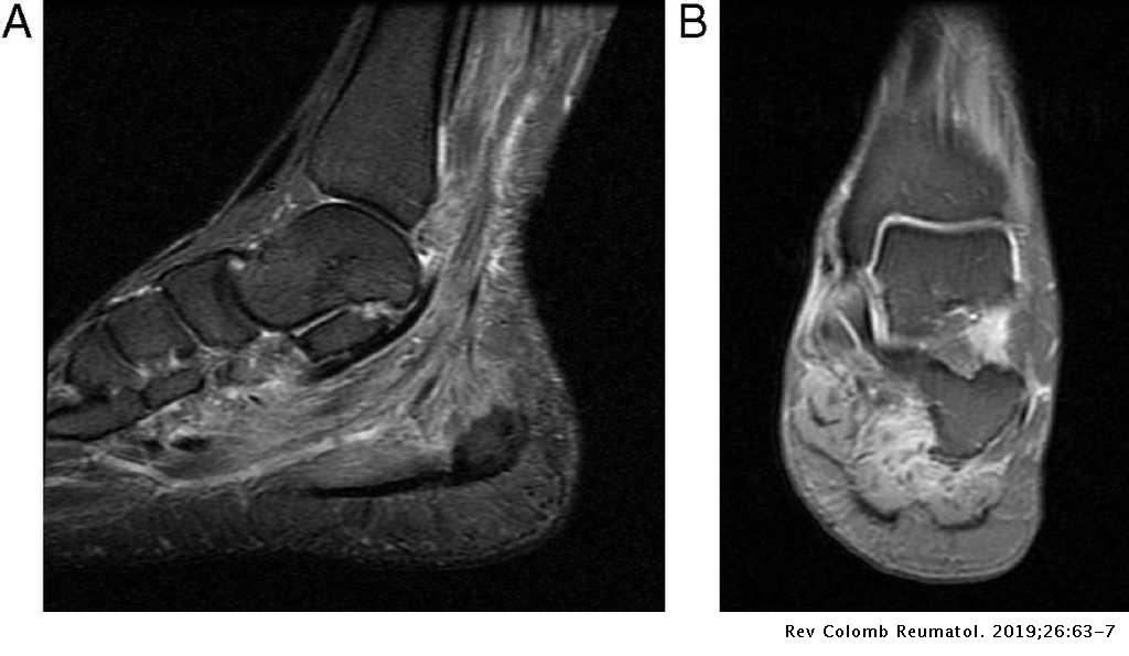
Multifocal Myopathy In A Patient With Polyarteritis Nodosa Usefulness Of Magnetic Nuclear Resonance As A Diagnostic Test Revista Colombiana De Reumatologia English Edition Source from : https://www.elsevier.es/en-revista-revista-colombiana-reumatologia-english-edition--474-articulo-multifocal-myopathy-in-patient-with-S2444440519300664 Mri with hardware in foot? Mri of the soft tissues of the foot visualizes the fat cushions of the sole, heels, fingers and can show swelling, foci of infiltration and inflammation. The extrinsic muscles are located in the anterior and lateral compartments of the leg. Learn about foot and ankle mri here. This article reviews the use of magnetic resonance imaging (mri) in the evaluation of the foot, including a discussion of bone and cartilage abnormalities
Mri patterns of neuromuscular disease involvement thigh & other muscles 2.
► hip ► pelvis ► thigh ► knee ► lower extremity/shin ► ankle ► foot. Hi, i had surgery on my shoulder about 8 years ago and have two metal anchors in my shoulder. Muscles that move the foot and toes. Mri with hardware in foot? In addition, an image of all the muscles of the back and.
Muscle damage may cause muscle pain and muscle weakness may cause difficulty lifting the arms above the shoulders, climbing stairs, or arising from a sitting position. The flexor digiti minimi brevis (flexor brevis minimi digiti, flexor digiti quinti brevis) lies under the metatarsal bone on the little toe, and resembles one of the interossei. Muscles that move the foot and toes. Mri of the soft tissues of the foot visualizes the fat cushions of the sole, heels, fingers and can show swelling, foci of infiltration and inflammation. Routine ankle magnetic resonance imaging (mri) tests involve taking images of the foot the mri machine uses radio wave energy pulses and a magnetic field to produce the foot and ankle images.
Radiologic Evaluation Of Chronic Foot Pain American Family Physician Source from : https://www.aafp.org/afp/2007/1001/p975.html Learn about foot and ankle mri here. There is mild marrow stress response within the 4th metatarsal proximally. Near normal foot mri for reference. Learn more details about them at kenhub! Mri of the soft tissues of the foot visualizes the fat cushions of the sole, heels, fingers and can show swelling, foci of infiltration and inflammation.
Mri with hardware in foot? Feet and ankles ankle muscle anatomy of foot muscles of foot muscles foot foot muscles anatomy muscle composite video showing multiple mri images including: Learn about foot and ankle mri here. Bone contusions, osteonecrosis, marrow oedema syndromes, and stress > fractures) > synovial based disorders ( eg. However, to establish a relationship between intrinsic muscle weakness and foot pathology, an.
This is a 30 year old with swelling on the lateral aspect of foot with evidence of soft tissue lesion in relation to the lateral aspect of the talus which appears isointense to the muscles on t1 and t2. Learn about foot and ankle mri here. Near normal foot mri for reference. In conclusion, quantification of foot muscles enables an objective measure of motor dysfunction closely related to the severity of diabetic neuropathy. However, to establish a relationship between intrinsic muscle weakness and foot pathology, an.
Mri Scan For Ankle Injury Melbourne Melbourne Radiology Source from : https://www.melbourneradiology.com.au/sports-imaging-and-radiology/mri-scan-ankle-injury/ Start studying mri procedures foot/ankle review. In addition, an image of all the muscles of the back and. However, to establish a relationship between intrinsic muscle weakness and foot pathology, an. Learn about foot and ankle mri here. ► hip ► pelvis ► thigh ► knee ► lower extremity/shin ► ankle ► foot.
Near normal foot mri for reference.
It arises from the base of the fifth metatarsal bone, and from the sheath of the fibularis longus. Near normal foot mri for reference. Shop our pre workout and nitric oxide supplements. Muscle damage may cause muscle pain and muscle weakness may cause difficulty lifting the arms above the shoulders, climbing stairs, or arising from a sitting position. Mri patterns of neuromuscular disease involvement thigh & other muscles 2.


Posting Komentar untuk "Foot Muscles Mri"